Brain tumor
Brain tumors are classified as primary, those that arise in the brain, or secondary, those that have spread to the brain from another part of the body. In the United States, about 24,000 people per year are diagnosed with a primary brain tumor. Primary brain tumors usually develop from glial cells, which provide the structural backbone of the brain and support the function of neurons. Most primary brain tumors in adults have no clear risk factors identified.
Brain tumors can produce symptoms due to local brain invasion, compression of healthy brain structures and by increasing pressure within the brain (increased intracranial pressure). Symptoms vary based on what parts of the brain are involved.
Cerebellar tonsillar ectopia/Chiari malformation
The cerebellum is the part of the brain that coordinates voluntary movements such as posture, balance, coordination, and speech, resulting in smooth and balanced muscular activity. Cerebellar tonsillar ectopia is when the cerebellar tonsils (bottom part of the cerebellum) extend into the spinal canal.
Cavernoma
A cavernoma, also known as a cavernous malformation, is a mass in the brain or spinal cord made up of abnormal dilated blood vessels. Cavernomas account for a large proportion (8-15%) of all brain and spinal vascular malformations. They are dynamic structures, changing in size and number over time. About a third of individuals with cavernomas develop symptoms, often between ages 20 - 40, which can include severe headache that is different from past headaches, nausea and vomiting, sensitivity to light, fainting, stroke-like symptoms, loss of consciousness and/or seizures.
Arachnoid cyst
The brain and spinal cord are covered by three protective membrane-linings called meninges. Sometimes, for unclear reasons, extra cerebral spinal fluid (CSF) can collect under the middle membrane - the arachnoid membrane. This leads to the formation of a benign (non-cancerous) collection of fluid called an arachnoid cyst. Arachnoid cysts account for approximately 1% of intracranial masses.
Arachnoid cysts usually are asymptomatic (do not present symptoms), but sometimes can cause headaches, neurologic deficits or seizures. For asymptomatic arachnoid cysts, management can include monitoring with regular brain imaging and neurologic examinations. Surgery is only indicated if symptoms develop.
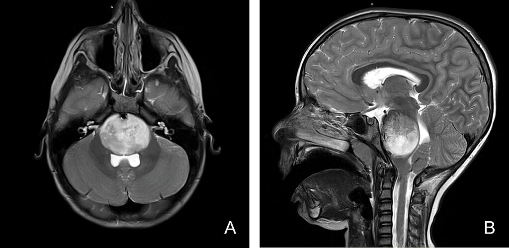
Cerebellopontine angle lesion
The cerebellopontine angle is a triangular space filled with cerebrospinal fluid (CSF). It is located in the posterior cranial fossa (the most posterior aspect of the skull base housing the brainstem and cerebellum).
Cerebellopontine angle (CPA) tumors are the most common neoplasms (abnormal growths) in the posterior fossa, accounting for 5-10% of intracranial tumors. Most CPA tumors are benign (non-cancerous), slow-growing tumors, with over 85% being vestibular schwannomas (acoustic neuromas), lipomas, vascular malformations, and hemangiomas. Symptoms may include headache, ringing in the ears, dizziness, hearing or visual changes and sensation changes in the face. Cerebellopontine angle tumors can cause nerve damage or compress the brain stem if not treated.
Chronic microvascular changes
Microvascular ischemic disease is a term that is used to describe changes to the small blood vessels in the brain. The cause of microvascular ischemic disease is not completely understood. It can be the result of plaque buildup and hardening (atherosclerosis) of the small blood vessels nourishing the brain. This is the same process that can narrow and damage heart blood vessels.